")
")


What happens during a brain dissection
At Institute Born-Bunge (IBB), every brain tells a story. For those affected by neurodegenerative diseases like Alzheimer’s, Parkinson’s, or ALS, that story may be complex and devastating, but through science, it can also become meaningful.
One of the most impactful ways to advance research into these conditions is through post-mortem brain donation. This process, while often not visible to the public, is a cornerstone of how we better understand the human brain and improve future diagnosis and treatment.

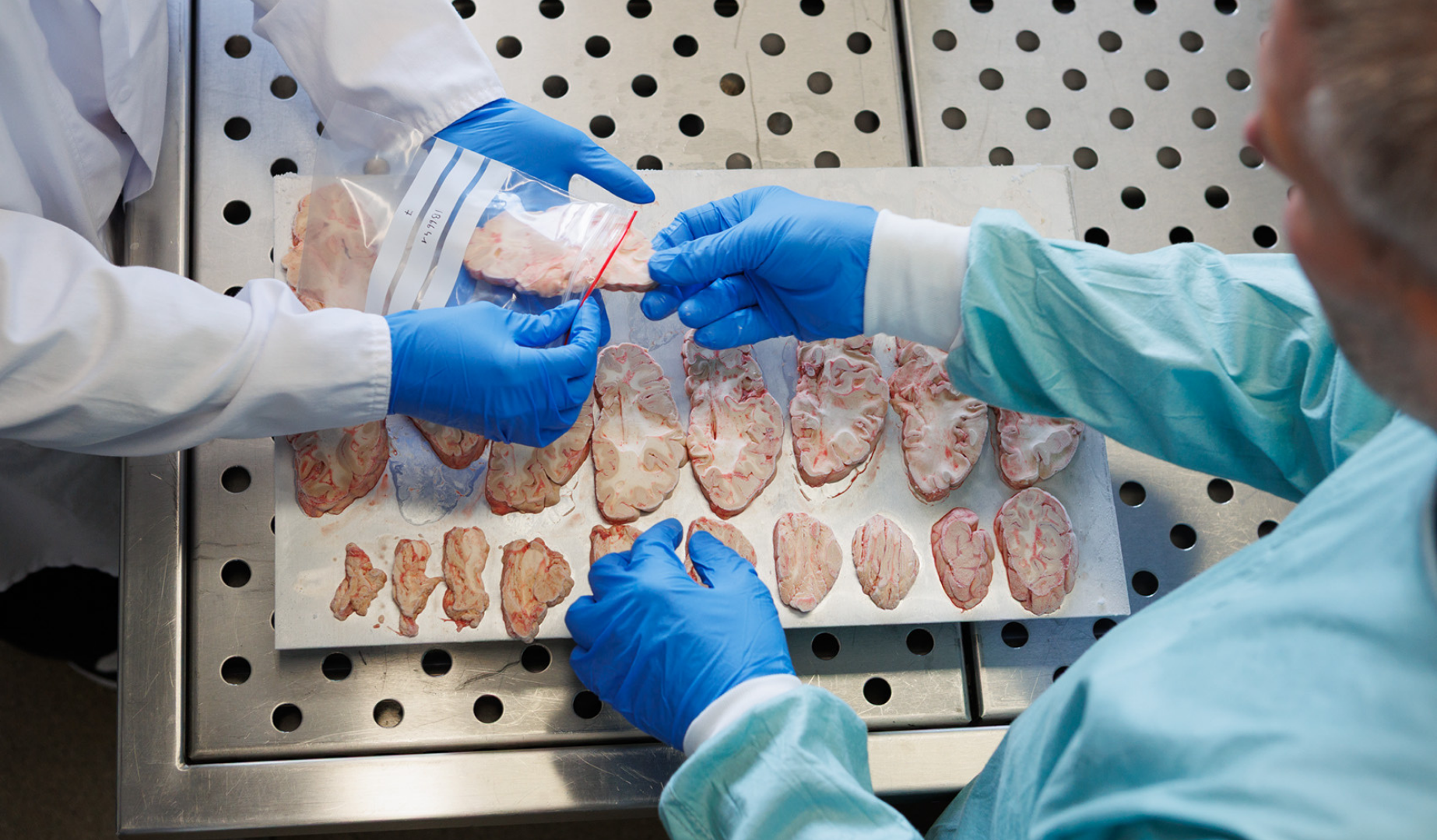
A race against the clock
When a person has agreed to donate their brain, time becomes critical. After death, a dedicated team steps in to begin the dissection within 12 hours. This rapid response is essential to preserve the tissue in a condition that makes meaningful scientific analysis possible. The brain is carefully removed and divided in two. One half is preserved in formalin, a solution that stabilizes the tissue for detailed visual and microscopic analysis. The other half is frozen immediately to preserve the molecular structure — allowing researchers to later study proteins, genes and other biological markers in their most natural state.
A closer look, one layer at a time
After a few weeks, the fixed half of the brain is photographed and examined. This is when neuropathologists begin a careful investigation. They select specific regions — such as the hippocampus, frontal cortex or brainstem — that are commonly affected in neurological diseases. These areas are then embedded in paraffin, thinly sliced, and placed on microscope slides for detailed staining. Different stains reveal different types of damage. Some highlight nerve cells, others myelin (the protective coating around nerves), and still others illuminate harmful proteins. In Alzheimer’s disease, for example, researchers use a silver stain to identify senile plaques, or antibodies to detect tau and amyloid proteins. In other diseases, like frontotemporal dementia or Parkinson’s, different markers are used to uncover telltale signs of degeneration.
A gift that keeps giving
Each donated brain becomes part of IBB’s NeuroBiobank, one of Europe’s most richly documented collections of brain tissue. This material is shared with researchers around the world to investigate causes, compare treatments, and develop earlier diagnostic tools. In fact, the way we classify many diseases today is directly shaped by what we’ve learned from these post-mortem examinations.